Echocardiography
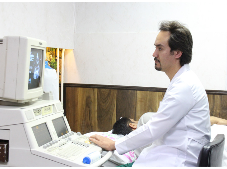
An echocardiography (Echo) is a graphic outline of the heart's movement. During an echo test, ultrasound (high-frequency sound waves) from a hand-held wand placed on your chest provides pictures of the heart's valves and chambers and helps the cardiologist evaluate the pumping action of the heart. Echo is often combined with Doppler ultrasound and color Doppler to evaluate blood flow across the heart's valves
The test is used to:
Pediatric echocardiography is ultrasonography used to evaluate the anatomical structure, simple to complex congenital defects, function of the heart and planning the time and kind of the surgery.
Stress echocardiogram is a noninvasive test used to assess the heart's response to stress or exercise and compare the results to when the heart is at rest.
The test is used to:
A dobutamine stress echocardiogram (DSE) may be used if you are unable to exercise. Dobutamine is put in a vein and causes the heart to beat faster. It mimics the effects of exercise on the heart, rest it is the same test as Exercise Stress Echo.